*vision:-
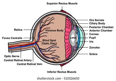
-eye -has three layers
:sclera- anteriorly called as cornea
-choroid
-retina-have receptors
-visual receptors(5 marks)
teloreceptors -stimulated from distance,stimulated by light,present more at fovea centralis
-cells:cone&rod
*rod:-
rod shaped
-less in number
-concentrated more toward periphery
-pigment present is rodopsin
-for dim light
*cone cell:-
coneshaped
-more in number for visual aequity about 120 million
-concentrated more toward fovea centralis
-pigments:-red-porphyropsin
green-iodopsin
blue-cynopsin
-bright light vision
*aqueous humor(5/2mark)
its atranspanrent liquid presemt in anterior diameter &posterior chamber infront of lens
-it is secreted in cilliary body and goes to posterior chamber through the pupil it enters into anterior chamber.
-it is drained at canal of schelmma which is at junction of sclera- corneal junction
-function:-provides nutritionand o2 to the avascular tissue lens & cornea
-maintains inraocular pressure within eyeball
n.v=16-20mmhg
if it is increased condition is called glaucoma which leads to detachmentwhich leads to blindness
maintains the shape of eyeball
*dark adaptation:-
-the subject is going to bright to dark room
-requires more than -20min
-resynthesis of rodopsin-retinol+opsin
-pupil dialation occurs
*light adaptation:-
subject going from dark room to bright light
-require less time-5
-cleavage of rodopsin-opsin& retinol
-pupil constriction occurs
*optic pathway:-(5marks)
 flow chart:-
resceptors-rods &cones
| 1st order neuron
bipolar cells
|
ganglionic cells
|
optic nerve
|
optic chiasma
|
optic tract
| 2nd order neuron
synapse in thalamus
|
optic cradiation
|3rd order neuron
visual cortex(17,18,19)
*refractive errors of vision:-
-myopia
-hypermyopia
-presbiopia
-astigmatism
*myopia
-can see nearby objects ,cant see far off objects
-anterior posterior dimensions of eyeball increases,light rays are focusses infront of retina
-correction by using bi-concave lens,it diverges the rays &focus on retina
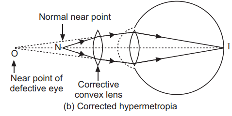
 *hypermetropia:-
can see far objects ,not near objects
-anterior posterior dimension of eyeball decreases,so light rays are focussed behind retina
-correction by using convex lens ,it converges the rays &focus on retina
 *presbyopia:
due to ageing (after 40 years )
-cause-due to weakening of cilliary muscles which hols lens
symptom:-can ssee far objects cant read read books
-correction-bifocal lens
*astigmatism:-
cause -corneal surface is not uniform or uneven
symptoms blurred vision
correction-cylindrical lens
*pupillary reflex:-
1)accomadation reflex
2)light reflex
1)accomadation reflex:-
human eye is set for distant region
- changes occur in eye during near vision is called accomadation reflex
-they are: medial rotation of eyebrows
constriction pupil
convexity of lens increases
flow chart:-
retina
|
opticnerve
|
optic tract
|
thalamus
|
visual cortex
|
visual motor neuron
|-
iii cranial nerve
|
medial rectus
|
cilliary nerve
*`light reflex-
beam of light thrown /focussed into the eye if constriction of pupil occurs then it is called direct light reflex
-if constriction of pupil occurs opposite eye is called indirect light reflex.
-significance:
-regulation of entry of light
-to diagnosis of encephalitis /neurosyphilis
pathway-
retina
|
optic nerve
|
optic chiasma
|
optic tract
pre tectal nucleaus in medulla
|
3rd cranial nerve(edinger westpal nuc.)
|
ciliary nerve
|
sphincter pupillar muscle
|
constriction of pupil

-eye -has three layers
:sclera- anteriorly called as cornea
-choroid
-retina-have receptors
-visual receptors(5 marks)
teloreceptors -stimulated from distance,stimulated by light,present more at fovea centralis
-cells:cone&rod
*rod:-
rod shaped
-less in number
-concentrated more toward periphery
-pigment present is rodopsin
-for dim light
*cone cell:-
coneshaped
-more in number for visual aequity about 120 million
-concentrated more toward fovea centralis
-pigments:-red-porphyropsin
green-iodopsin
blue-cynopsin
-bright light vision
*aqueous humor(5/2mark)
its atranspanrent liquid presemt in anterior diameter &posterior chamber infront of lens
-it is secreted in cilliary body and goes to posterior chamber through the pupil it enters into anterior chamber.
-it is drained at canal of schelmma which is at junction of sclera- corneal junction
-function:-provides nutritionand o2 to the avascular tissue lens & cornea
-maintains inraocular pressure within eyeball
n.v=16-20mmhg
if it is increased condition is called glaucoma which leads to detachmentwhich leads to blindness
maintains the shape of eyeball
*dark adaptation:-
-the subject is going to bright to dark room
-requires more than -20min
-resynthesis of rodopsin-retinol+opsin
-pupil dialation occurs
*light adaptation:-
subject going from dark room to bright light
-require less time-5
-cleavage of rodopsin-opsin& retinol
-pupil constriction occurs
*optic pathway:-(5marks)
resceptors-rods &cones
| 1st order neuron
bipolar cells
|
ganglionic cells
|
optic nerve
|
optic chiasma
|
optic tract
| 2nd order neuron
synapse in thalamus
|
optic cradiation
|3rd order neuron
visual cortex(17,18,19)
*refractive errors of vision:-
-myopia
-hypermyopia
-presbiopia
-astigmatism
*myopia
-can see nearby objects ,cant see far off objects
-anterior posterior dimensions of eyeball increases,light rays are focusses infront of retina
-correction by using bi-concave lens,it diverges the rays &focus on retina
can see far objects ,not near objects
-anterior posterior dimension of eyeball decreases,so light rays are focussed behind retina
-correction by using convex lens ,it converges the rays &focus on retina
due to ageing (after 40 years )
-cause-due to weakening of cilliary muscles which hols lens
symptom:-can ssee far objects cant read read books
-correction-bifocal lens
*astigmatism:-
cause -corneal surface is not uniform or uneven
symptoms blurred vision
correction-cylindrical lens
*pupillary reflex:-
1)accomadation reflex
2)light reflex
1)accomadation reflex:-
human eye is set for distant region
- changes occur in eye during near vision is called accomadation reflex
-they are: medial rotation of eyebrows
constriction pupil
convexity of lens increases
flow chart:-
retina
|
opticnerve
|
optic tract
|
thalamus
|
visual cortex
|
visual motor neuron
|-
iii cranial nerve
|
medial rectus
|
cilliary nerve
*`light reflex-
beam of light thrown /focussed into the eye if constriction of pupil occurs then it is called direct light reflex
-if constriction of pupil occurs opposite eye is called indirect light reflex.
-significance:
-regulation of entry of light
-to diagnosis of encephalitis /neurosyphilis
pathway-
retina
|
optic nerve
|
optic chiasma
|
optic tract
pre tectal nucleaus in medulla
|
3rd cranial nerve(edinger westpal nuc.)
|
ciliary nerve
|
sphincter pupillar muscle
|
constriction of pupil

note:-constriction of pupil occurs on both sides becuase fibers from PTN(PRE TECTAL NUC.)pass to both sides to edingar westpal nuc. of 3rd cranial nerve get excited & constricted of pupil occur on both sides
*argyl robertsons pupil:-
accomadation reflex is present ,light reflex is absent becuase there is lesion in pretectal nucleas that is the center of light reflex
EG:neurosyphilis
*visual acuity:-its the resolving power of eye to different 2 points
test:-1)snellers chart:-meant for distant region (6 METERS)
2)JAEGARS CHART:-meant for near region(30 cm)
colour vision:-
unable to appreciate primary colours(blue,green,red)
it is a sex linked disorder females are carriers male are suffers)
Test:wool matching test
edridge green lantern test
Comments